Notes Chapter 18 Body Fluids and Circulation
Class 11 students can refer to Chapter 18 Body Fluids and Circulation notes given below which is an important chapter in the class 11 Biology book. These notes and important questions and answers have been prepared based on the latest CBSE and NCERT syllabus and books issued for the current academic year. Our team of Biology teachers has prepared these notes for class 11 Biology for the benefit of students so that you can read these revision notes and understand each topic carefully.
Body Fluids and Circulation Notes Class 11 Biology
Refer to the notes and important questions given below for Body Fluids and Circulation which is really useful and has been recommended by Class 11 Biology teachers. Understanding the concepts in detail and then solving questions by yourself will help you to learn all topics given in your NCERT Books.
BODY FLUIDS AND CIRCULATION
Body fluids are the medium of transport of nutrients, oxygen and other important substances in the body.
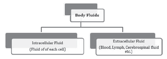
Blood is the most commonly used body fluid in most of the higher organisms. Lymph also transports certain substances like protein and fats.
Blood
Blood is a fluid connective tissue composed of a fluid matrix, plasma and the blood corpuscles. It forms about 30-35% of the extracellular fluid. It is slightly alkaline fluid having pH7.4.
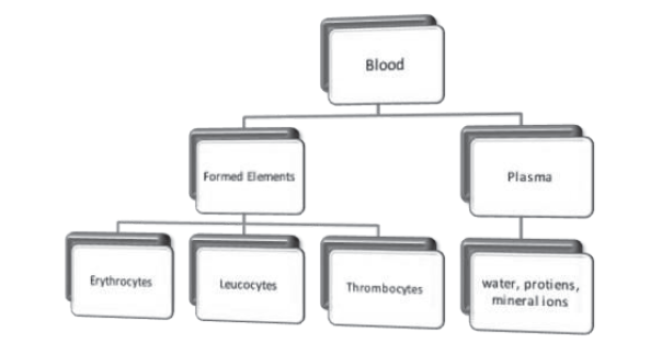
- Plasma is straw coloured viscous fluid that constitutes 55% of blood volume. It consists of 90-92% water, 6-8% protein (fibrinogens, albumins and globulins), glucose,
amino acids and small amount of minerals like Na+, Ca++, Cl- etc. - Erythrocytes, leucocytes and platelets are collectively called formed elements.
- Erythrocytes are most abundant cells in human body. Total blood count of RBCs is 5- 5.5 million, which is slightly less in females due to menstruation. It is formed in bone marrow. Nucleus is absent in mammalian RBCs having biconcave shape.
- Every 100 ml of blood contain 12-16 gm. of haemoglobin. They have life span of 120 days. They are destroyed in spleen( graveyard of RBCs)
- Leucocytes or WBCs are colourless due to absence of haemoglobin. 6000-8000 of WBCs are present in each ml. of blood.
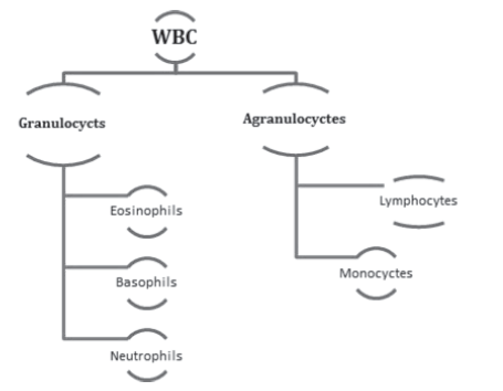
- Neutrophils are most abundant and basophils are least abundant WBCs. Monocytes and neutrophils are phagocytic cells which destroy foreign organisms.

- Basophils secrete histamine, serotonin and heparin that are involved in inflammatory reactions.
- Eosinophils resist infection and allergic reactions. B and T lymphocytes are responsible for immune response of the body.
Thrombocytes or platelets are cell fragments produced from megakaryocytes in bone marrow. 150000-350000 platelets are present in each ml of blood. Platelets are involved in
clotting or coagulation of blood in case of injuries.
Blood Groups – blood of human beings differ in certain aspects although it appear same in all individuals. Two main types of grouping are ABO and Rh.
ABO grouping is based on presence or absence of two surface antigens RBC, antigen A and antigen B. The plasma of an individual also contains two antibodies produced in response of
antigens.

- During blood transfusion, blood of donor has to be matched with blood of recipients to avoid clumping of RBCs.
- Group ‘O’ blood can be donated to any individual with any blood group, so it is called universal donor.
- Person with ‘AB’ blood group can receive blood from any person of any group, so it is called universal recipient.
Rh grouping – Rh antigen (similar to Rhesus monkey) are observed on surface of RBCs of majority of individuals (about 80%). Such people are called Rh positive (Rh+) and those in
whom this antigen is absent are called Rh negative (Rh– ).
- Erythroblastosis foetalis- if father blood is Rh+ and mother blood is Rh–, the foetus blood is Rh+. During the delivery of first child there is a possibility of exposure of mother blood with foetus blood to develop antibodies in mother blood. In subsequent pregnancy the mother’s blood can leak into foetus blood and destroy the foetus RBC.
This case is called erythroblastosis foetalis.
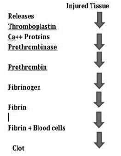
Coagulation of blood (Blood Clotting)
When an injury is caused to a blood vessel bleeding starts which is stopped by a process called blood clotting. An injury or trauma stimulates the platelets in the blood to release
certain factors that activate the mechanism of coagulation. Calcium play important role in blood clotting.
Lymph
During flow of blood through capillaries, some water soluble substances move out in the space between cells of tissues. This fluid released out is called interstitial fluid or tissue fluid.
It is similar to the blood but has fewer blood proteins, less calcium and phosphorus and high glucose concentration.
- It is a colourless fluid containing specialized lymphocytes that provide immune response to body.
- Main function of lymph is to provide immunity, carry proteins and fats molecules and transport oxygen, food materials, hormones etc.
Circulatory Pathways
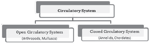
- All vertebrates have a muscular chambered heart.
Fish – 2 chambered heart
Amphibian and Reptiles (except crocodile) – 3 chambered heart.
Crocodile, Birds and Mammals – 4 chambered heart.
Human Circulatory System – consists of 4 chambered muscular heart, closed branching blood vessels and circulatory fluid blood.
Heart is the mesodermally derived muscular organ, present in thoracic cavity between the two lungs protected by double membrane of pericardium.
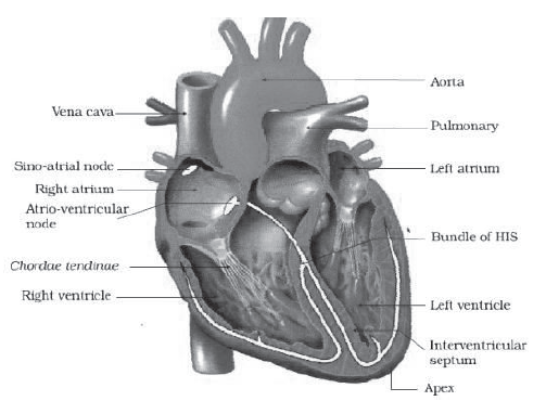
- The upper two chamber is called atria and lower two chambers are called ventricles.
Interatrial septum separate the right and left atrium and thick walled inter ventricle septum separate the ventricles. - The opening between right atrium and right ventricle is guarded by a three muscular flaps called tricuspid valve. Bicuspid or mitral valve guards the left atrium and ventricle.
- The opening of right and left ventricle to pulmonary artery and aorta respectively is controlled by semilunar valve.
- The nodal tissue present on upper right corner of right atrium is called SAN (sinoatrial node) and those on lower left corner of right atrium is called AVN ( atrioventricular node).
- The purkinje fibres along with right and left bundles form the bundle of HIS. The nodal musculature has ability to generate action potential.
- SAN generate maximum number of action potential and is responsible for rhythmic contraction of heart. Therefore it is called pace maker.
Cardiac Cycle
To begin with, all four chambers are in relaxed state called joint diastole. As the bicuspid and tricuspid valves are open, blood from pulmonary vein and vena cava flows to left and right ventricle respectively. Semilunar valves are closed at this stage.
SA node generates action potential that contracts both atria (atrial systole). The action potential passes to AV node and bundle of HIS transmit it to ventricular musculature to cause ventricular systole. At the same time atria undergoes relaxation diastole to close the bicuspid and tricuspid valve.
Semilunar valves open into circulatory system that relax the ventricle and close the valves to prevent back flow of blood.
As the pressure inside ventricle decreases the bicuspid and tricuspid valve open to repeat the process or cardiac cycle.
During each cardiac cycle two sounds are produced. The first sound (lub) is due to closure of bicuspid and tricuspid valve and 2nd heart sound (dub) is due to closure of semilunar valve.
ECG (Electrocardiograph) is a graphical representation of electrical activity of heart during cardiac cycle. The electrocardiograph machine is used to obtain electrocardiogram. The patient is connected to three electrical leads to wrists and left ankle.
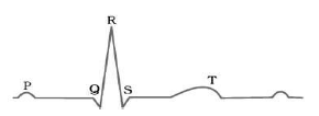
- The P-wave represents the electrical excitation of atria (depolarisation) which leads to contraction of atria.
- The QRS-wave represents the depolarisation of ventricles, which initiates the ventricular contraction.
- The T-wave represents the return of ventricle from exited to normal state (repolarization). The end of T-wave marks the end of systole. Counting the number of QRS complex in given period of time determine the heartbeat rate.
Double Circulation
Flow of same blood twice through the heart once in oxygenated form and other in deoxygenated form is called double circulation. It includes systematic and pulmonary circulation.
Systematic circulation includes flow of oxygenated blood from the left ventricle to all parts of body and deoxygenated blood from various body parts to the right atrium. All systematic circulation starts form aorta and ends at superior vena cava, inferior vena cava or coronary sinus to right atrium.
The systematic circulation provides oxygen, nutrients and other substances to the tissues and take CO2 and other harmful substances away for removal.
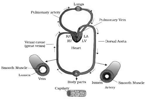
Pulmonary Circulation
The flow of deoxygenated blood from the right ventricle to the lungs and the return of oxygenated blood from the lung to the left atrium is called pulmonary circulation.
Two pulmonary veins from each lung transport the oxygenated blood to the left atrium.
Double circulation prevents the mixing of oxygenated and deoxygenated blood.
Regulation of Cardiac Activity
- Normal activities of heart are regulated by nodal tissue (SA and AV node), so the heart is myogenic.
- A special neural centre in medulla oblongata moderates the cardiac function by ANS.Sympathetic nerve can increase the rate of heart beat and parasympathetic nerve of ANS decrease the rate of heart beat.
- Adrenal medullary hormone also increases the cardiac output.
Disorder of Circulatory System
1. Hypertension (high blood pressure) – Blood pressure higher than (120/80) . 120 mm Hg is the systolic that is pumping pressure and 80 mm Hg is the diastole, resting pressure. It leads to heart disease and affect vital organs like brain and kidney.
2. Coronary Artery Disease (CAD)- commonly called atherosclerosis that affects the blood vessels that supply blood to heart muscles due to deposition of fat, calcium, cholesterol that makes the arteries lumen narrower.
3. Angina- also called angina pectoris, acute chest pain due to less supply of oxygen to heart muscles. It may occur in elderly male and female. It occurs due to restricted blood flow.
4. Heart failure- heart does not pump enough blood to meet the requirement of body. It is also known as congestive heart failure because congestion of lung is one of its causes.Heart failure is different from heart attack ( heart muscle is damaged by inadequate blood supply) and cardiac arrest ( when heart stops beating).
5. Coronary Thrombosis- formation of clot in the coronary artery is coronary thrombosis. It occurs most frequently in the left anterior descending coronary artery.