Notes Chapter 20 Locomotion and Movement
Class 11 students can refer to Chapter 20 Locomotion and Movement notes given below which is an important chapter in the class 11 Biology book. These notes and important questions and answers have been prepared based on the latest CBSE and NCERT syllabus and books issued for the current academic year. Our team of Biology teachers has prepared these notes for class 11 Biology for the benefit of students so that you can read these revision notes and understand each topic carefully.
Locomotion and Movement Notes Class 11 Biology
Refer to the notes and important questions given below for Locomotion and Movement which is really useful and has been recommended by Class 11 Biology teachers. Understanding the concepts in detail and then solving questions by yourself will help you to learn all topics given in your NCERT Books.
LOCOMOTION AND MOVEMENT
Locomotion is the voluntary movement of an individual from one place to another.Walking, running, climbing, swimming are the examples of locomotion. All locomotion are movement but all movements are not locomotion.
Types of Movement
Cells of the human body show three main types of movements:
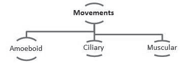
- Macrophages and leucocytes in blood exhibit amoeboid movements. Coordinated movement of cilia in trachea to remove dusts particles and passage of ova through fallopian tube is example of Ciliary movements.
- Movement of limbs, jaw, tongue, etc. need muscular movement. Contractile property of muscles is used in movement in higher organism including human beings.
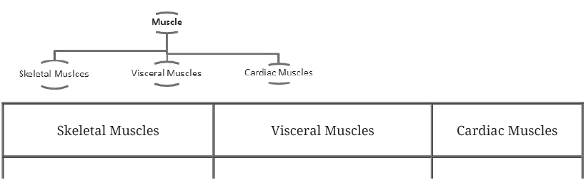
Muscles are specialized tissues of mesodermal origin. They have property like excitability, contractility, extensibility and elasticity.
Based on their location, three types of muscles are identified

Skeletal Muscle is made up of muscles bundles (fascicles), held together by collagenous connective tissue called fascia.

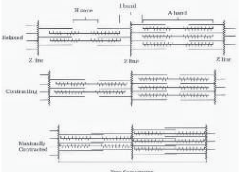
- Each muscle bundle contains a number of muscle fibres. Each muscle fibre is lined by plasma membrane called sarcolemma enclosing sarcoplasm. Partially arranged myofibrils are present in muscle bundle having alternate light and dark bands due to presence of protein- actin and myosin
- Light bands contain actin and is called I-band (isotropic band) and dark band contains myosin, called A-band (anisotropic band). Both bands are present parallel to each other in longitudinal fashion.
- In centre of each I-band is elastic fibre called ‘Z’ line. In the middle of A-band is thin fibrous ‘M’ line. The portion of myofibrils between two successive ‘Z’ lines is the functional unit of contraction called a sarcomere.
- At resting stage thin filament overlaps the thick filament. The part of thick filament not overlapped is called ‘H’ zone.
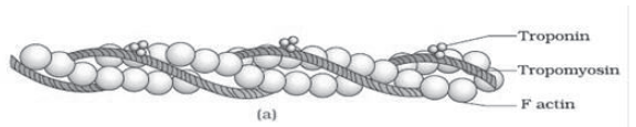
Structure of contractile Protein
Each thin filament (actin) is made of two ‘F’ actins helically wounded to each other. Two filaments of another protein, tropomyosin runs close to it. A complex protein Troponin is distributed at regular intervals on the tropomyosin.
Each myosin filament is made of many monomeric proteins called Meromyosins. Each meromyosin has globular head with short arm and tails. Globular head has ATP binding sites.
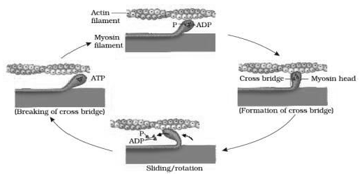
Mechanism of muscle contraction
- The mechanism of muscle contraction is explained by sliding mechanism theory in which thin filament slide over thick filament.
- Muscle contraction start with signal sent by CNS via motor neuron. Neural signal release neurotransmitter ( Acetyl choline) to generate action potential in the sarcolemma.
- This causes the release of Ca ++ from sarcoplasmic reticulum.
- Ca ++ activates actin which binds to the myosin head to form a cross bridge.
- These cross bridges pull the actin filaments causing them to slide over the myosin filaments and thereby causing contraction.
- Ca ++ are then returned to sarcoplasmic reticulum which inactivate the actin. Cross bridges are broken and the muscles relax.
Muscles are classified as:
Red fibres (aerobic muscles-) contain myoglobin that has plenty of mitochondria to use large amount of oxygen stored in them.
White fibres-the muscle fibres containing less number of myoglobin are called white fibres.
Skeletal System
Framework of bones and cartilage forms the skeletal system. In human beings, it consists of 206 bones and some cartilages. The two principle division of skeletal system are:
1. Axial Skeleton (80 bones)- includes skull, vertebral column, sternum and ribs constitute axial system.
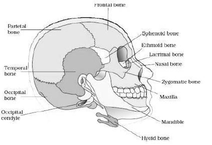
- The skull (22 bones) is composed of cranial and facial bones. Cranial (8 bones) forms
- protective covering for brain (cranium). The facial region consists of 14 skeletal systems that form front part of skull. Hyoid bone (U-shaped) forms the base of buccal cavity.
- The middle ear bone (Malleus, Incus and Stapes) collectively called Ear Ossicles. Skull joins with vertebral column with two occipital condyle.
- Vertebral column consists of 26 serially arranged vertebrae. First vertebra is atlas that combines with occipital condyle. Other includes Cervical-7, thoracic -12, lumbar -5, sacral – 1 coccoygeal -1.
- 12 pairs of ribs connected dorsally to vertebral column and ventrally to sternum. 11th and 12th rib bones are not connected with sternum and are called floating ribs.
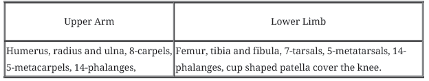
Appendicular Skeleton- includes bones of limbs and girdles. Each limb contains 30 bones.
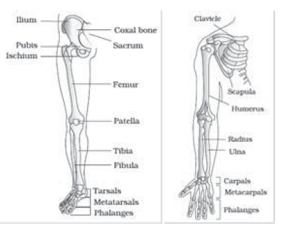
Pectoral and Pelvic girdle bones help in the articulation of the upper and the lower limbs respectively with the axial skeleton.
Pectoral girdle consists of a clavicle and a scapula.
Pelvic girdle consists of two coxal bones. Each coxal bone is formed by the fusion of three bones – ilium, ischium and pubis.
Joints – are points of contact between bones, or between bones and cartilage.
1. Fibrous joints- do not allow any movements. Present in flat skull bones to form cranium.
2. Cartilaginous joints- bones are held together with the help of cartilage present in vertebrae. Permits limited movements.
3. Synovial joints- fluid filled synovial cavity, provide considerable movements. Ball and socket joint, hinge joints, pivot joints, gliding joints etc.
Disorders of Muscular and Skeletal System
Myasthenia gravis- auto immune disorder affecting neuromuscular junction causing fatigue, weakening and paralysis of skeletal system.
Muscular Dystrophy- degeneration of skeletal muscles due to genetic disorder.
Osteoporosis – decreased bone mass in old age leading to chance of fracture due to decreased estrogen.
Arthritis- inflammation of joints.
Gout- inflammation of joints due to accumulation of uric acid crystals.
Tetany- Rapid spasms in muscle due to low Ca ++ in body fluid